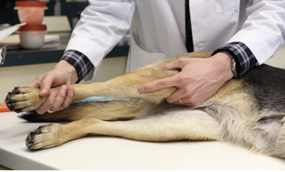
Cruciate Disease
Cranial Cruciate Ligament Disease (CCL)
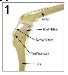
In animals, the knee is known as the stifle. The stifle consists of two long bones called the Femur and Tibia. The knee cap or Patella sits within the patellar ligament and is attached to the tibia by the patellar tendon. The area where the patellar tendon is attached is called the Tibial Tuberosity. The top of the tibia is called the Tibial Plateau. Between the two bones, there is a cushion called the Meniscus.
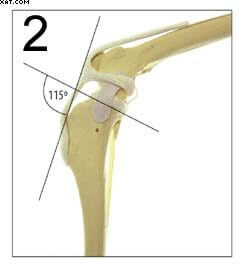
During weight bearing the femur slides down the tibial plateau. The cranial cruciate ligament (CCL or ACL in humans) stops this downward slide. The CCL is in constant tension during weight bearing. This biomechanical process can result in rupture of the ligament. This entire process is directly related to the angle between the tibial plateau slope and the patellar tendon. A more perpendicular angle results in a more stable joint.
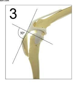
The TTA involves an osteotomy (or cut) in the non-weight bearing portion of the tibia, just behind the tibial tuberosity. The tibial tuberosity is advanced to achieve a perpendicular relationship between the tibial plateau slope and the patellar tendon. This relationship results in a stable joint.
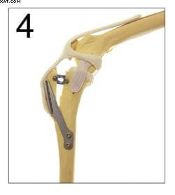
The advanced tibial tuberosity is secured using titanium implants. Healing takes about 12 weeks in most dogs. Most dogs, however, are walking on the limb the day after surgery. Implants do not require removal unless there are complications. If you have questions please call us at (810) 653-3988 or (810) 412-0336.
Explore Our Complete List of Veterinary Services in Davison, MI
- Echocardiograms
- Orthopedic Surgery
- Pain Management
- Parasite Screening & Preventatives
- Ultrasounds
- Dermatology
- Ophthalmology
- X-Ray & Radiology
- Nutritional
- Allergy Testing & Treatment
- Laser Therapy
- Dental Care
- Surgery
- Microchipping
- Senior Wellness
- Vaccinations
- Spay & Neuter
- In-House Laboratory
- Wellness Exams
